

Knowing the differences between an MRI and X-ray can help you make an informed decision with your doctor about which imaging test is right for your condition.
What is an X-Ray?
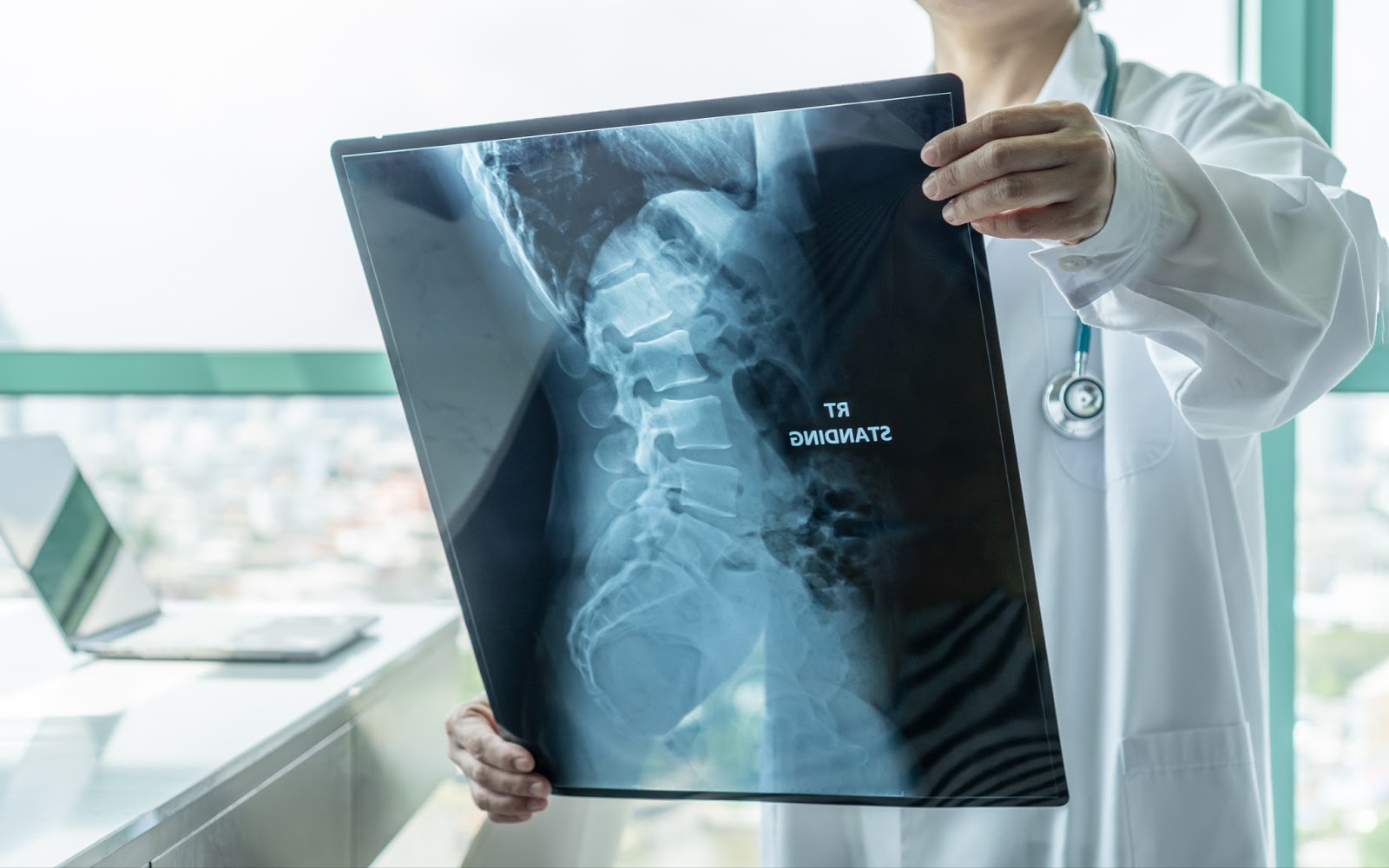
X-rays are a form of electromagnetic radiation similar to radio waves that are used to produce images of internal tissues, bones and organs. Areas within the body with high levels of calcium, such as bones and teeth, block the radiation, which is what causes them to appear white on the image. Soft tissues in the body, such as blood, skin, fat and muscle, allow the radiation to pass through and appear dark gray on the image.X-rays are most commonly used to look at the bones and joints in the body, but sometimes are used to detect problems affecting soft tissue. Problems that may be detected during an X-ray include:
- Bone fractures
- Dislocations
- Narrowed joint spaces
- Scoliosis
- Bone spurs
What is an MRI (Magnetic Resonance Imaging)?
Magnetic resonance imaging (MRI) is a non-invasive imaging technology that uses radio waves to produce images of body structures. An MRI scanner is a long cylinder with a narrow tube in the center; a moveable bed slides inside the tube for scanning.MRIs use magnets and radio frequencies to create images of body structures. To better explain how the technology works, it’s important to consider the human body’s structure: The human body is made up of six main elements, including oxygen, carbon, hydrogen, nitrogen, calcium, and phosphor. And roughly 60% of the body is water.
Every water molecule in the body is like a tiny magnet. When the water molecules are exposed to the magnetic field generated by an MRI scan, it causes these molecules to line up and spin at a particular speed. A secondary magnetic field turns the molecules to face new directions and once it switches off, the molecules realign, allowing the MRI scanner to peek inside the body and create an image.
MRIs are commonly used to investigate soft tissue injuries or conditions and are useful for detecting musculoskeletal conditions, including:
- Cartilage loss
- Spinal injuries
- Joint inflammation
- Torn or detached ligaments, tendons, muscles and cartilage
- Tumors
- Inflammatory bowel disease
- Brain injuries
- Developmental anomalies
- Multiple sclerosis
- Dementia
- Infection
- And more.
 Accredited Business. Rating A+ Click for Review