Overview

An acromioclavicular joint separation, or AC separation, is a very frequent injury among physically active people. In this injury the clavicle (collar bone) separates from the scapula (shoulder blade). It is commonly caused by a fall directly on the "point" of the shoulder or a direct blow received in a contact sport. Football players and cyclists who fall over the handlebars are often subject to AC separations.

In general, most AC injuries don't require surgery. There are certain situations, however, in which surgery may be necessary. Most patients recover with full function of the shoulder. The period of disability and discomfort ranges from a few days to 12 weeks depending on the severity of the separation. Disruption of the AC joint results in pain and instability in the entire shoulder and arm. The pain is most severe when the patient attempts overhead movements or tries to sleep on the affected side.

What does the inside of the shoulder look like?

The shoulder is the most mobile joint in the human body, with a complex arrangement of structures working together to provide the movement necessary for daily life. Unfortunately, this great mobility comes at the expense of stability. Several bones and a network of soft tissue structures (ligaments, tendons, and muscles), work together to produce shoulder movement. They interact to keep the joint in place while it moves through extreme ranges of motion. Each of these structures makes an important contribution to shoulder movement and stability. Certain work or sports activities can put great demands upon the shoulder, and injury can occur when the limits of movement are exceeded and/or the individual structures are overloaded.Click here to read more about shoulder structure.

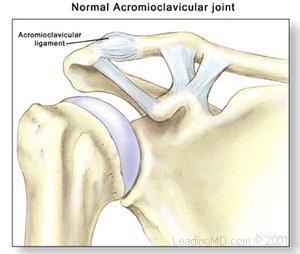
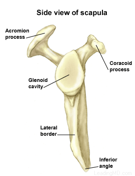
What is an AC joint separation?

An AC joint separation, often called a shoulder separation, is a dislocation of the clavicle from the acromion. This injury is usually caused by a blow to the shoulder, or a fall in which the individual lands directly on the shoulder or an outstretched arm. AC joint separations are most common in contact sports, such as football and hockey.

The severity of an acromioclavicular joint injury depends on which supporting structures are damaged, and the extent of that damage. Tearing of the acromioclavicular ligament alone is not a serious injury, but when the coracoclavicular ligaments are ruptured, the whole shoulder unit is involved, thus complicating the dislocation.

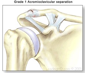
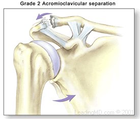
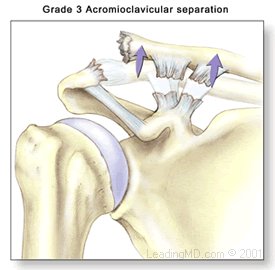
Simple AC injuries are classified in three grades ranging from a mild dislocation to a complete separation:

Grade I - A slight displacement of the joint. The acromioclavicular ligament may be stretched or partially torn. This is the most common type of injury to the AC joint.

Grade II - A partial dislocation of the joint in which there may be some displacement that may not be obvious during a physical examination. The acromioclavicular ligament is completely torn, while the coracoclavicular ligaments remain intact. 

Grade III - A complete separation of the joint. The acromioclavicular ligament, the coracoclavicular ligaments, and the capsule surrounding the joint are torn. Usually, the displacement is obvious on clinical exam. Without any ligament support, the shoulder falls under the weight of the arm and the clavicle is pushed up, causing a bump on the shoulder. 

There are a total of six grades of severity of AC separations. Grades I-III are the most common. Grades IV-VI are very uncommon and are usually the result of a very high-energy injury such as one that might occur in a motor vehicle accident. Grades IV-VI are all treated surgically because of the severe disruption of all the ligamentous support for the arm and shoulder.

Shoulder Structure

Bones and Joints

The bones of the shoulder:

  • The humerus is the upper arm bone. This is the "ball" of the shoulder's "ball and socket" joint.
  • The scapula is the flat, triangular bone commonly called the shoulder blade. Prominent areas of the scapula serve as attachment points for many muscles and ligaments.
    • The glenoid is the shallow "socket" on the side of the scapula that receives the 'ball' of the humerus. Together they form the "ball and socket" arrangement of the shoulder.
    • The scapular spine is a horizontal ridge along the back of the scapula that divides the scapula into upper and lower regions.
    • The acromion is the end of the scapular spine. It projects up to form the top of the shoulder.
    • The coracoid process is a projection towards the front of the scapula and is an attachment site for several muscles and ligaments.
  • The clavicle is the collarbone. Although it appears to be straight, it actually forms an S-shape when seen from above.
  • The thorax or rib cage, is an anchor for several muscles and ligaments. Although the ribs do not physically attach to the scapula, the thorax stabilizes and maintains proper positioning of the scapula so that the arm can function to its fullest capacity.

Additionally, there are four bone junctions, or joints:

  • The glenohumeral joint is the main joint of the shoulder. Here, the glenoid on the scapula and the head of the humerus come together. The fairly flat socket of the glenoid surrounds only 20% - 30% of the humeral head. Because of its poor fit, this joint relies heavily on the surrounding soft tissue for support. The labrum, a ring of fibrocartilage tissue, attaches to the glenoid and deepens the socket to encircle more of the humerus.
  • The acromioclavicular joint, or AC joint, is the bony point on the top of the shoulder. It stabilizes the scapula to the chest, by connecting the acromion on the scapula to the clavicle, or "collarbone". A thick disk of fibrocartilage acts as a shock absorber between the two bones. The surrounding capsule and ligaments give this joint great stability.
  • The sternoclavicular joint, or SC joint, connects the other end of the clavicle to the sternum, or "breastbone". Like the AC joint, this joint contains a fibrocartilage disk that helps the bones achieve a better fit. It also gets excellent support from its joint capsule and surrounding ligaments.
  • The scapulothoracic articulation is the area where the scapula, embedded in muscle, glides over the thoracic rib cage. The surrounding muscles and ligaments keep the scapula properly positioned so that the arm can move correctly.

Cartilage

There are two types of cartilage in the shoulder:

  • Articular cartilage is the shiny white coating that covers the end of the humeral head and lines the inside surface of the glenoid. It has two purposes:
    • To provide a smooth, slick surface for easy movement
    • To be a shock absorber and protect the underlying bone
  • Fibrocartilage is the thick tissue that forms the disks of the AC and SC joints and the labrum, the ring that deepens the glenoid. Fibrocartilage has three roles:
    • To act as a cushion in shock absorption
    • To help stabilize the joint by improving the fit of the bones
    • To act as a spacer and improve contact between the articular cartilage surfaces

Ligaments

The shoulder relies heavily on ligaments for support. Ligaments attach bone to bone and provide the "static" stability in a joint. Ligaments will alternately become tight and loose with normal motion. They keep the joint within the normal limits of movement.

  • The glenohumeral ligaments attach in layers from the glenoid labrum to form the joint capsule around the head of the humerus.
  • The coracoacromial arch is the group of ligaments that spans the bony projections of the coracoid process and the acromion.
  • The coracoclavicular ligaments and the acromioclavicular ligament provide most of the support for the AC joint.

Muscles and Tendons

Muscles and tendons work together in the shoulder to provide the "dynamic" stability of the shoulder.

There are four muscle groups in the shoulder:

  • The rotator cuff muscles are the subscapularis, the supraspinatus, the infraspinatus, and the teres minor. They are the primary stabilizers that hold the "ball" of the humerus to the glenoid "socket". The socket is too shallow to offer much security for the humerus. These four muscles form a "cuff" around the humeral head, securing it firmly in the socket. As its name implies, this group of muscles also rotates the arm. The rotator cuff protects the glenohumeral joint from dislocation, allowing the large muscles that control the shoulder to power the arm with great mobility.
  • The biceps tendon complex also helps keep the humeral head in the glenoid and helps raise the arm.
  • The scapulothoracic muscles attach the scapula to the thorax. Their main function is to stabilize the scapula to allow for proper shoulder motion.
  • The superficial muscles of the shoulder are the large, powerful outer layer of muscles that are important to the overall function of the shoulder. This group includes the deltoid muscle, which covers the rotator cuff muscles.

Bursae

A bursa is a pillow-like sac filled with a small amount of fluid. Bursae (plural) reduce friction and allow smooth gliding between two firm structures, like bone and tendon or bone and muscle. There are over 50 bursae in the human body; the largest is the subacromial bursa (under the acromion) in the shoulder. The subacromial bursa and the subdeltoid bursa (under the deltoid muscle) are often considered as one structure. This bursa separates the rotator cuff and the deltoid muscle, from the acromion.

Symptoms

What are the signs and symptoms of an AC joint separation?

Grade I Injury:

  • There may be tenderness at the joint when touched.
  • There may be some bruising around joint.
  • There is minor pain with arm movement.
  • There is no pain in the area of the coracoclavicular ligaments.


Grade II Injury:

  • There is moderate to severe pain at the joint.
  • Swelling may be present.
  • There is pain with arm movement.
  • There may be a small bump on the top of the shoulder where the clavicle ends.
  • The clavicle may move when pushed.
  • The area of the coracoclavicular ligaments may be painful when touched.


Grade III Injury:

  • Typically, the injured person immediately supports the elbow while holding the arm close to the side.
  • This prevents the pain outward arm movement would cause.
  • Pain is present with any arm motion.
  • Pain is present around the joint and in the area of the coracoclavicular ligaments.
  • Swelling is present.
  • As the joint moves, a popping sound may occur.
  • There is shoulder deformity and a bump on the top of the shoulder.
  • The AC joint is very unstable.


Diagnosis

How is an acromioclavicular separation diagnosed?

After evaluating the history of the patient's injury the doctor will examine the shoulder area looking for signs of fracture or dislocation by comparing the overall position of the arm and shoulder to the uninjured side. The AC joint itself is easily examined because it is located right under the skin. The doctor will gently feel the bones and soft tissue around the joint and between the acromion and clavicle. There may be a bump, tenderness or instability, which would suggest a joint separation. Crepitus (noise) as the bones move may indicate a fracture. The doctor also evaluates the patient's range of motion and performs tests to isolate specific areas of pain and weakness.

Further evaluation may include:

  • A diagnostic anesthetic injection, which blocks pain, can help confirm the diagnosis.
  • X-rays can confirm a diagnosis of an AC joint separation and can help the physician determine whether the injury is a separation or a fracture. A new cross body X-ray has been recently developed which is very helpful in confirming the diagnosis and may be in general use in the near future.
Treatment

How is an acromioclavicular separation treated?

The treatment of an AC separation depends on the grade of the injury. The classification helps the physician choose the correct treatment approach. Grades I - III are usually treated non-operatively. The vast majority of patients will have a period of discomfort. Once this discomfort disappears, the shoulder is usually fully functional, although the patient may still have a minor cosmetic defect at the injury site.

Some patients with grade III AC separations may be candidates for early surgical reconstruction. A discussion between the physician and patient should focus on the patient's expectations and possible return to sports. Many surgeons prefer to first treat the AC separation conservatively. If grade III patients develop problems or do not heal properly surgical reconstruction is an option. (Conservative and surgical treatment for grades I - III have essentially the same results after 1 year.)

Non-Operative Treatment

Most Grade I - III AC separations are treated successfully with non-operative treatment that may include:

  • ice to reduce pain and swelling.
  • rest and a protective sling until pain subsides. This usually takes about 1-2 weeks.
  • pain and anti-inflammatory medications.
  • A rehabilitation program to restore normal motion and strength is begun as soon as tolerated with gentle exercises and progresses as healing allows.

Depending on the grade of injury, most patients heal within 2 to 3 months without surgical intervention. The patient is allowed to return to sports when there is full and painless range of motion, no more tenderness when the AC joint is touched, and manual traction does not cause pain. This usually takes about 2 weeks for a grade I injury, 6 weeks for a grade II injury, and up to 12 weeks for a grade III injury.

Operative Treatment

Surgery may be necessary for AC separations that do not respond well to non-operative treatment. If, after 2 to 3 months, pain continues in the AC joint with overhead activity or in contact sports, surgery may be necessary. There are some physicians who offer early surgery for a select group of Grade III AC separations based upon the activities and demands these patients place upon the shoulder.

These patients include:

  • young, active individuals (over the age of 13).
  • laborers whose jobs require heavy overhead work.
  • athletes in non-contact sports whose overhead movements are stressful and frequent.

A variety of surgical methods have been used to stabilize a separated AC joint. The surgical technique most often performed involves the reconstruction of the coracoclavicular ligaments and the excising (removal) of the distal (shoulder) end of the clavicle. Distal clavicle resection without the repair of the ligaments may lead to excessive rotation of the scapula. Reconstruction studies show that the AC joint can be adequately stabilized by:

  • a fixation across the acromioclavicular space with pins or plates.
  • loop fixation from the clavicle to the coracoid process using synthetic materials.
  • The most common reconstructive procedures today use a screw or suture loop to stabilize the joint.

In a distal clavicle resection, about 10-15mm of the clavicle is removed through a two-inch incision above the joint. The AC ligament is then transferred from the bottom of the acromion into the cut end of the clavicle to replace the torn ligament.

What types of complications may occur?

Complications of AC joint injuries are persistent instability of the shoulder girdle or residual pain with activity. These complications can be present with either non-operative treatment or operative treatment. Failure of the acromioclavicular ligament and coracoclavicular ligaments to heal can lead to pain and a sense of instability with overhead activity. If the end of the clavicle remains unstable because of lack of scarring, contact sports or overhead tasks may be painful.

Other complications associated with the reconstruction of the AC or CC ligaments are related to hardware failure. Fixation of the clavicle to the coracoid process is difficult because of the rotation of the clavicle with all overhead activity. The screws used to fix these two bones together can pull out if the patient does not wear a sling after surgery as instructed.

Most surgeons today will securely fix the clavicle to the coracoid with dissolvable sutures or with a screw that is removed at about three months.

Recovery

Non-Operative

Patients with lower energy AC joint injuries that respond to conservative non-operative treatment can recover in as little as one week for a Grade I injury to an average of twelve weeks for a Grade III injury.

Operative

Specific recovery programs following surgical reconstruction of the AC joint vary depending on the type of surgery performed. General care recommendations include:

  • The use of a sling is required for about 4 weeks.
  • Incisions must be kept dry for about three days after surgery.
  • Stitches are usually removed 7 - 10 days after surgery.
  • Lower arm exercises may begin immediately. With an open surgical technique the arm may be immobilized for 2-3 weeks before motion exercises are begun.
  • No lifting for 3 weeks.
  • Overhead motion is limited for the first 8-12 weeks because the fixation of the clavicle to the coracoid process prevents the normal rotation necessary for overhead movements.
  • The patient is allowed use of the arm at waist height to write or perform computer work for the first 12 weeks. After that time, the screw (if used) is removed, or the sutures have softened enough to begin more aggressive range of motion and strengthening exercises.
  • After the removal of the sling, 6-8 weeks of physical therapy is necessary to regain full motion.
  • Strength and velocity motions of the throwing athlete may be delayed for 4-6 months.
FAQs

Do all AC separations require surgery?

No. In fact the vast majority of AC separations do very well with conservative treatment of the symptoms. Most AC injuries are grade I, II, or III and these generally do not require surgery. Usually the joint remains sore for two to six weeks and then full return to activity is the norm. Only unstable grade III injuries and high-energy AC separations, which are often the result of motor vehicle accidents, require surgery for full recovery.

Will the "bump" ever go away?

The clavicle will become stable in its newly elevated position, but without surgery the "bump" will remain. The joint will function normally and will not remain tender to touch or movement. This minor cosmetic deformity will persist but will not interfere with overhead activities or participation in sports.

Are there downsides to a resection of the AC joint?

An AC resection is a procedure in which the end of the clavicle is removed and the acromioclavicular ligament in reattached into the end of the clavicle to replace the ligament torn during injury. Once the initial injury has healed and the clavicle has regained stability from scar tissue there is no functional loss with an AC resection. In the rare instance that the AC joint remains painful after a separation, but does not require stabilization, an AC resection is very effective in relieving pain without sacrificing function. If, however, the clavicle is unstable at the time of resection, a full reconstruction of the coracoclavicular ligaments is necessary to maintain the stability of the upper extremity.

Will I be able to return to athletics if an AC injury is not treated?

Absolutely. Most athletes in contact sports have had a low energy AC separation at some time in their careers. Except for the slight deformity that remains, there is no clinical significance to a healed AC separation. Occasionally high-energy AC separations that have disruption of the AC and CC ligaments will require surgery, but these injuries are usually apparent early on with a correct X-ray evaluation. Grade I, II, and most grade III AC separations will heal without treatment and a full return to sports can be expected

References

1. Rockwood and Green Textbook of Fractures for a complete description of AC injuries and their classification.

2. Richards RR. Acromialclavicular injuries. Instr Course Lectures 42:259-269,1993.

3. Basmania CJ. Acromioclavicular Joint Injuries in Athletes. In: Garrett WE, Speer KP, Kirkendall DT, eds. Principles and Practice of Orthopaedic Sports Medicine. Philadelphia, PA: Lippincott Williams and Wilkins; 2000:511-534.

4. Soslowsky LJ, Carpenter JE, Kuhn JE. Biomechanics of the Shoulder. In: Garrett WE, Speer KP, Kirkendall DT, eds. Principles and Practice of Orthopaedic Sports Medicine. Philadelphia, PA: Lippincott Williams and Wilkins; 2000:367-372.

5. Pasque CB, McGinnis DW, Yurko-Griffin L. Shoulder. In: Sullivan JA, Anderson SJ, eds. Care of the Young Athlete. American Academy of Orthopaedic Surgeons and American Academy of Pediatrics; 2000;323-327.

6. Thompson WO, Warren RF, Barnes RP, Hunt S. Shoulder Injuries. In: Schenck, Jr, RC ed. Athletic Training and Sports Medicine. 3rd edition. Rosemont, IL: American Academy of Orthopaedic Surgeons; 1999:220-232.

Accredited Business.  Rating A+  Click for Review