Overview

Labral tears of the shoulder have been a particular focus of attention since the advent of the arthroscope, a small instrument that allows the orthopaedic surgeon to clearly see inside the shoulder joint and view the labrum, its environment, and any injuries that may have occurred. The arthroscope, in conjunction with anatomic dissections, history, physical examination, and symptoms has allowed orthopaedic surgeons to better understand and treat a variety of injuries to the labrum.

What does the inside of the shoulder look like?

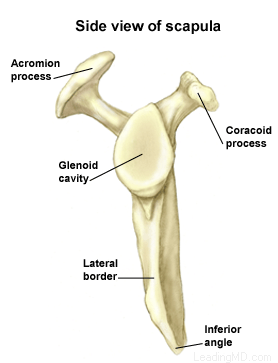
The shoulder is the most mobile joint in the human body. It consists of a complex arrangement of structures working together to provide the movement necessary for daily life. Unfortunately, this great mobility comes at the expense of stability. Four bones and a network of soft tissue structures (ligaments, tendons, and muscles) work together to produce shoulder movement. They interact to keep the joint in place while it moves through extreme ranges of motion. Each of these structures makes an important contribution to shoulder movement and stability. Certain work or sports activities can put great demands upon the shoulder, and injury can occur when the limits of movement are exceeded and/or the individual structures are overloaded. Click here to read more about shoulder structure.

What is the labrum and what does it do?

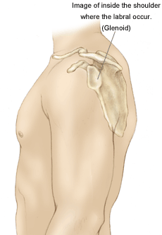
The labrum is a disk of cartilage on the glenoid, or "socket" side of the shoulder joint. The labrum helps stabilize the joint and acts as a "bumper" to limit excessive motion of the humerus, the "ball" side of the shoulder joint. More importantly, it holds the humerus securely to the glenoid, almost as if suction were involved. Although the glenoid itself is a relatively flat surface, the labrum's cuff-like contour gives the glenoid a more concave shape. The secure but flexible fit of the humerus within the glenoid permits the great range motion of the healthy shoulder.

How is the labrum injured?

Tears of the labrum can be the result of:

  • a direct fall or blow to the arm resulting in abnormal movement of the humerus.
  • repetitive trauma of the greater tuberosity and rotator cuff on the posterior (back of) labrum. This is termed internal impingement (pinching of the soft tissues) and is most commonly seen in baseball and tennis athletes whose arms are frequently in overhead positions.

A very common labral injury is a tear that occurs on the top of the labrum, extending from the front to the back of the cartilage. This is known as a SLAP tear ("SLAP" is an acronym for superior labral anterior to posterior tear). This injury affects the attachment of the biceps tendon to the glenoid. An injury in this area can be extremely painful, and can cause the biceps tendon to rupture.

At least five types of SLAP tears have been identified. Treatment depends on the stability of the biceps anchor and the type of tear that has occurred. These injuries are commonly the result of:

  • a fall on an outstretched arm.
  • a forceful lifting maneuver.
  • repetitive throwing.

It is very important for the surgeon to determine whether or not the labral tear is associated with instability of the shoulder. A tear of the labrum not associated with instability can be treated surgically by itself, but the treatment of a labral tear in an unstable shoulder will only be successful if the shoulder is surgically stabilized at the same time.

Shoulder Structure

Bones and Joints

The bones of the shoulder:

  • The humerus is the upper arm bone. This is the "ball" of the shoulder's "ball and socket" joint.
  • The scapula is the flat, triangular bone commonly called the shoulder blade. Prominent areas of the scapula serve as attachment points for many muscles and ligaments.
    • The glenoid is the shallow "socket" on the side of the scapula that receives the 'ball' of the humerus. Together they form the "ball and socket" arrangement of the shoulder.
    • The scapular spine is a horizontal ridge along the back of the scapula that divides the scapula into upper and lower regions.
    • The acromion is the end of the scapular spine. It projects up to form the top of the shoulder.
    • The coracoid process is a projection towards the front of the scapula and is an attachment site for several muscles and ligaments.
  • The clavicle is the collarbone. Although it appears to be straight, it actually forms an S-shape when seen from above.
  • The thorax or rib cage, is an anchor for several muscles and ligaments. Although the ribs do not physically attach to the scapula, the thorax stabilizes and maintains proper positioning of the scapula so that the arm can function to its fullest capacity.

Additionally, there are four bone junctions, or joints:

  • The glenohumeral joint is the main joint of the shoulder. Here, the glenoid on the scapula and the head of the humerus come together. The fairly flat socket of the glenoid surrounds only 20% - 30% of the humeral head. Because of its poor fit, this joint relies heavily on the surrounding soft tissue for support. The labrum, a ring of fibrocartilage tissue, attaches to the glenoid and deepens the socket to encircle more of the humerus.
  • The acromioclavicular joint, or AC joint, is the bony point on the top of the shoulder. It stabilizes the scapula to the chest, by connecting the acromion on the scapula to the clavicle, or "collarbone". A thick disk of fibrocartilage acts as a shock absorber between the two bones. The surrounding capsule and ligaments give this joint great stability.
  • The sternoclavicular joint, or SC joint, connects the other end of the clavicle to the sternum, or "breastbone". Like the AC joint, this joint contains a fibrocartilage disk that helps the bones achieve a better fit. It also gets excellent support from its joint capsule and surrounding ligaments.
  • The scapulothoracic articulation is the area where the scapula, embedded in muscle, glides over the thoracic rib cage. The surrounding muscles and ligaments keep the scapula properly positioned so that the arm can move correctly.

Cartilage

There are two types of cartilage in the shoulder:

  • Articular cartilage is the shiny white coating that covers the end of the humeral head and lines the inside surface of the glenoid. It has two purposes:
    • To provide a smooth, slick surface for easy movement
    • To be a shock absorber and protect the underlying bone
  • Fibrocartilage is the thick tissue that forms the disks of the AC and SC joints and the labrum, the ring that deepens the glenoid. Fibrocartilage has three roles:
    • To act as a cushion in shock absorption
    • To help stabilize the joint by improving the fit of the bones
    • To act as a spacer and improve contact between the articular cartilage surfaces

Ligaments

The shoulder relies heavily on ligaments for support. Ligaments attach bone to bone and provide the "static" stability in a joint. Ligaments will alternately become tight and loose with normal motion. They keep the joint within the normal limits of movement.

  • The glenohumeral ligaments attach in layers from the glenoid labrum to form the joint capsule around the head of the humerus.
  • The coracoacromial arch is the group of ligaments that spans the bony projections of the coracoid process and the acromion.
  • The coracoclavicular ligaments and the acromioclavicular ligament provide most of the support for the AC joint.

Muscles and Tendons

Muscles and tendons work together in the shoulder to provide the "dynamic" stability of the shoulder.

There are four muscle groups in the shoulder:

  • The rotator cuff muscles are the subscapularis, the supraspinatus, the infraspinatus, and the teres minor. They are the primary stabilizers that hold the "ball" of the humerus to the glenoid "socket". The socket is too shallow to offer much security for the humerus. These four muscles form a "cuff" around the humeral head, securing it firmly in the socket. As its name implies, this group of muscles also rotates the arm. The rotator cuff protects the glenohumeral joint from dislocation, allowing the large muscles that control the shoulder to power the arm with great mobility.
  • The biceps tendon complex also helps keep the humeral head in the glenoid and helps raise the arm.
  • The scapulothoracic muscles attach the scapula to the thorax. Their main function is to stabilize the scapula to allow for proper shoulder motion.
  • The superficial muscles of the shoulder are the large, powerful outer layer of muscles that are important to the overall function of the shoulder. This group includes the deltoid muscle, which covers the rotator cuff muscles.

Bursae

A bursa is a pillow-like sac filled with a small amount of fluid. Bursae (plural) reduce friction and allow smooth gliding between two firm structures, like bone and tendon or bone and muscle. There are over 50 bursae in the human body; the largest is the subacromial bursa (under the acromion) in the shoulder. The subacromial bursa and the subdeltoid bursa (under the deltoid muscle) are often considered as one structure. This bursa separates the rotator cuff and the deltoid muscle, from the acromion.

Symptoms

What are the signs and symptoms of a labral tear?

The primary symptom of a labral tear that is not associated with instability is pain in the area of the injury. Patients often say:

  • the pain is located at the back on top of the shoulder or in the front on top of the shoulder.
  • the pain feels like it is deep inside.
  • palpation (pressing) on the shoulder does not duplicate the pain that they have.

Labral tears that involve the biceps tendon (SLAP tears) can make using the biceps tendon painful. Overhead activity with inward rotation of the shoulder (which occurs when pitching or serving a tennis ball) causes pain. In addition, using a tool like a screwdriver can cause pain because the biceps is the primary muscle used for this motion.

Patients with labral tears from internal impingement (a pinching of the soft tissues) will complain that they have pain with throwing. Usually the pain is worst when the arm changes direction from late cocking to forward acceleration. In the shoulder, this pain is located posterior (behind) and superior (above). If there is a related partial thickness rotator cuff tear, there may also be lateral (on the side) pain.

Diagnosis

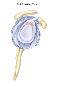
The physician will first take a history of the patient's injury in order to learn how it occurred. As mentioned earlier, the pain from a labral tear is located at the site of the injury, and is usually referred to as being deep inside the shoulder. A thorough physical exam may reveal other sources of injury or another disorder. During a physical examination of the shoulder:

  • the physician will measure motion to reveal any stiffness, which can be a sign of a labral tear.
  • an instability exam will be performed.
  • the O'Brien's test which focuses on the biceps and reveals pain, can help diagnose a labral tear.

X-rays will rule out other problems such as arthritis, fracture, impingement, or malignancy.

The confirming test for a labral tear is an MRI preceded by an arthrogram. This procedure greatly enhances the diagnostic accuracy by allowing tears of the labrum to be seen more clearly.

Treatment

Non-operative Treatment for Labral Tears

There are probably a large number of untreated labral tears that heal spontaneously since the labrum has a rich blood supply that helps the healing process (except in the anterior and superior locations).

Physical therapy is helpful for certain cases such as internal impingement, in which the rotator cuff rubs the posterior labrum. Physical therapy options include:

  • changing the throwing mechanism.
  • strengthening muscles that externally rotate the shoulder.
  • stretching the posterior capsule with the help of a physical therapist.

Operative Treatment for Labral Tears

Tears that need treatment are usually seen in the chronic stage when spontaneous healing has not occurred. Tears of the biceps tendon anchor are unlikely to heal without treatment because the biceps constantly pulls on the labrum. Chronic tears may require surgery if patients are unwilling or unable to modify their activities sufficiently to allow for healing and comfort. Surgery should be considered if:

  • pain is unresponsive to anti-inflammatory medications such as ibuprofen, rest, and activity modification.
  • the labrum has not healed after an acute injury.

The arthroscope allows direct visualization and evaluation of the entire shoulder joint. Using this small instrument, the orthopaedic surgeon first examines the labrum and all attachment sites for instability requiring repair. Once other disorders are treated or ruled out, the labral injury is addressed.

Arthroscopic treatment is the standard of practice for most labral injuries not associated with instability. The majority of these can be treated with simple debridement (removal of abnormal, damaged, or excess tissue). This procedure will eliminate flaps that may impinge the movement of the humerus on the glenoid and/or remove any unstable portion of the labrum.

Certain painful and unstable SLAP tears, in which the biceps is detached, need special attention. If the biceps tendon anchor is no longer firmly attached to the glenoid, it must be re-attached to the bone. The surgeon uses suture anchors and /or arthroscopic knot tying techniques to repair the torn labrum of younger patients.

It is often safer for older individuals to have a debridement of the labrum and a biceps tenodesis. A tenodesis is a procedure in which the biceps is repaired to the bone outside the glenohumeral joint to relieve pain, yet allow it to retain some strength and function. This procedure can be a good choice for older patients because the blood supply to the labrum diminishes with age, making the healing process more difficult. The decision to perform a tenodesis repair should be made by the doctor and patient together, after a thorough discussion of the surgical options and the healing process.

What types of complications can occur?

Complications associated with surgery for labral tears are few:

  • The risk of infection in arthroscopic procedures is extremely low and can be a complication of any surgical procedure.
  • Stiffness after surgery is unlikely since debridement allows early arm movement.
  • Physical therapy will usually relieve stiffness resulting from the sling that is needed for four or more weeks for labral repairs.
  • Failure of healing can occur due to technique, biology, or the patient's unwillingness to follow post-operative instructions and the prescribed rehabilitation program.
Recovery

In the early recovery period, physical therapy to regain hand and elbow range of motion can begin immediately. Labral tears that require only debridement are allowed early range of motion and physical therapy to prevent stiffness.

  • Sutures are removed seven to ten days after surgery and showering can resume.
  • Shoulder range of motion exercises after a biceps repair begin approximately six weeks after surgery, when the healing process is complete.

Physical therapy concentrates on treating factors that may have caused the labral injury, such as poor throwing mechanics. The patient's strength and rotator cuff health can be maximized with therapy, which helps the shoulder recover from what may have been a long period of inactivity due to pain.

Labral injuries that involve repair or tenodesis of the biceps tendon require a period of immobilization to allow the tendon to heal back to bone. After approximately four weeks of sling immobilization, physical therapy may begin.

Overhead athletes with SLAP repairs can expect a substantial delay before they can resume throwing. It usually takes about six months for the patient to regain full endurance and throwing velocity.

FAQs

Will a labral tear heal without treatment?

There are no good natural history studies on labral injuries. There are acute (sudden) labral tear injuries that are likely to heal without surgery. In chronic (longstanding) cases, however, there are no successful non-operative treatments.

What are the causes of a SLAP tear?

The causes of SLAP tears are a subject of debate among orthopaedic surgeons. There are several injury patterns that can lead to a tear. The most common causes are thought to be:

  • a fall on the outstretched hand that drives the humerus upward and causes the superior labrum to tear.
  • a sudden and often unexpected load applied to the biceps, which can cause a tear.
  • extremes of external rotation and abduction (movement away from the body) during throwing that causes the labrum to "peel back" from its attachment.

Why would my surgeon perform a tenodesis rather than a repair?

The decision to perform a tenodesis is based upon the location of the tear, the amount of biceps involved, and the quality of the remaining tendon. The more degenerative the tissue, the more likely it is that a tenodesis will be a successful treatment. The patient's age is a secondary concern, but is a factor (along with the other variables) that should be considered when assessing the likelihood of success of a primary repair versus the results obtainable with a tenodesis.

References

1.Labral Lesions Karzel, Snyder Operative Arthroscopy, Chapter 45 Second Edition edited by J.B. McGinty, R.B. Caspari, R.W. Jackson, G.C. Poehling. Lippencott-Raven Publishing, Philadelphia; 1996.

2.Cooper DE, et al. Anatomy, histology, and vascularity of the glenoid labrum: an anatomical study. JBJS. 1992;74:46-52.

3. Andrews JR, Carson WG. The arthroscopic treatment of glenoid labrum tears-the throwing athlete. Orthop Trans. 1984;8:44.

4. Snyder SJ et al. SLAP lesions of the shoulder. Arthroscopy. 1990;6:274-279. 5. Snyder SJ et al. MRI arthrography of the shoulder a new imaging technique. Clin Sports Med. 1993;1:123-136.

5.Snyder SJ et al. MRI arthrography of the shoulder a new imaging technique. Clin Sports Med. 1993;1:123-136.

6. Altcheck DW et al. Arthroscopic labral debridement: a three year follow up study. AJSM. 1992;20:702-706.

Accredited Business.  Rating A+  Click for Review