Overview

Frozen shoulder, or adhesive capsulitis, is a condition that begins with a gradual onset of pain and a limitation of shoulder motion. The discomfort and loss of movement can become so severe that even simple daily activities become difficult. Although much is known about this condition, there continues to be considerable controversy about its causes and the best ways to treat it.

What does the inside of the shoulder look like?

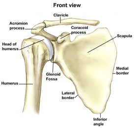
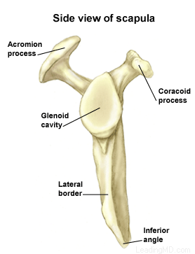
The shoulder is the most mobile joint in the human body, with a complex arrangement of structures working together to provide the movement necessary for daily life. Unfortunately, this great mobility comes at the expense of stability. Several bones and a network of soft tissue structures (ligaments, tendons, and muscles), work together to produce shoulder movement. They interact to keep the joint in place while it moves through extreme ranges of motion. Each of these structures makes an important contribution to shoulder movement and stability. Certain work or sports activities can put great demands upon the shoulder, and injury can occur when the limits of movement are exceeded and/or the individual structures are overloaded. Click here to read more about shoulder structure.

What is frozen shoulder?

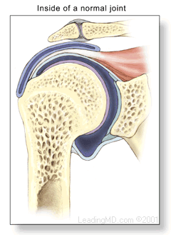
Frozen shoulder, also called adhesive capsulitis, is a thickening and tightening of the soft tissue capsule that surrounds the glenohumeral joint, the ball and socket joint of the shoulder. When the capsule becomes inflamed, scarring occurs and adhesions are formed. This scar formation greatly intrudes upon the space needed for movement inside the joint. Pain and severely limited motion often occur as the result of the tightening of capsular tissue.

There are two types of frozen shoulder: primary adhesive capsulitis and secondary adhesive capsulitis.

  • Primary adhesive capsulitis is a subject of much debate. The specific causes of this condition are not yet known. Possible causes include changes in the immune system, or biochemical and hormonal imbalances. Diseases such as diabetes mellitus, and some cardiovascular and neurological disorders may also be contributing factors. In fact, patients with diabetes have a three times higher risk of developing adhesive capsulitis than the general population. Primary adhesive capsulitis may affect both shoulders (although this may not happen at the same time) and may be resistant to most forms of treatment.
  • Secondary (or acquired) adhesive capsulitis develops from a known cause, such as stiffness following a shoulder injury, surgery, or a prolonged period of immobilization. 

With no treatment, the condition tends to last from one to three years. Many patients are unwilling to endure the pain and limitations of this problem while waiting for it to run its natural course. Even after many years, some patients will continue to have some stiffness, but no serious pain or functional limitations.

Shoulder Structure

Bones and Joints

The bones of the shoulder:

  • The humerus is the upper arm bone. This is the "ball" of the shoulder's "ball and socket" joint.
  • The scapula is the flat, triangular bone commonly called the shoulder blade. Prominent areas of the scapula serve as attachment points for many muscles and ligaments.
    • The glenoid is the shallow "socket" on the side of the scapula that receives the 'ball' of the humerus. Together they form the "ball and socket" arrangement of the shoulder.
    • The scapular spine is a horizontal ridge along the back of the scapula that divides the scapula into upper and lower regions.
    • The acromion is the end of the scapular spine. It projects up to form the top of the shoulder.
    • The coracoid process is a projection towards the front of the scapula and is an attachment site for several muscles and ligaments.
  • The clavicle is the collarbone. Although it appears to be straight, it actually forms an S-shape when seen from above.
  • The thorax or rib cage, is an anchor for several muscles and ligaments. Although the ribs do not physically attach to the scapula, the thorax stabilizes and maintains proper positioning of the scapula so that the arm can function to its fullest capacity.

Additionally, there are four bone junctions, or joints:

  • The glenohumeral joint is the main joint of the shoulder. Here, the glenoid on the scapula and the head of the humerus come together. The fairly flat socket of the glenoid surrounds only 20% - 30% of the humeral head. Because of its poor fit, this joint relies heavily on the surrounding soft tissue for support. The labrum, a ring of fibrocartilage tissue, attaches to the glenoid and deepens the socket to encircle more of the humerus.
  • The acromioclavicular joint, or AC joint, is the bony point on the top of the shoulder. It stabilizes the scapula to the chest, by connecting the acromion on the scapula to the clavicle, or "collarbone". A thick disk of fibrocartilage acts as a shock absorber between the two bones. The surrounding capsule and ligaments give this joint great stability.
  • The sternoclavicular joint, or SC joint, connects the other end of the clavicle to the sternum, or "breastbone". Like the AC joint, this joint contains a fibrocartilage disk that helps the bones achieve a better fit. It also gets excellent support from its joint capsule and surrounding ligaments.
  • The scapulothoracic articulation is the area where the scapula, embedded in muscle, glides over the thoracic rib cage. The surrounding muscles and ligaments keep the scapula properly positioned so that the arm can move correctly.

Cartilage

There are two types of cartilage in the shoulder:

  • Articular cartilage is the shiny white coating that covers the end of the humeral head and lines the inside surface of the glenoid. It has two purposes:
    • To provide a smooth, slick surface for easy movement
    • To be a shock absorber and protect the underlying bone
  • Fibrocartilage is the thick tissue that forms the disks of the AC and SC joints and the labrum, the ring that deepens the glenoid. Fibrocartilage has three roles:
    • To act as a cushion in shock absorption
    • To help stabilize the joint by improving the fit of the bones
    • To act as a spacer and improve contact between the articular cartilage surfaces

Ligaments

The shoulder relies heavily on ligaments for support. Ligaments attach bone to bone and provide the "static" stability in a joint. Ligaments will alternately become tight and loose with normal motion. They keep the joint within the normal limits of movement.

  • The glenohumeral ligaments attach in layers from the glenoid labrum to form the joint capsule around the head of the humerus.
  • The coracoacromial arch is the group of ligaments that spans the bony projections of the coracoid process and the acromion.
  • The coracoclavicular ligaments and the acromioclavicular ligament provide most of the support for the AC joint.

Muscles and Tendons

Muscles and tendons work together in the shoulder to provide the "dynamic" stability of the shoulder.

There are four muscle groups in the shoulder:

  • The rotator cuff muscles are the subscapularis, the supraspinatus, the infraspinatus, and the teres minor. They are the primary stabilizers that hold the "ball" of the humerus to the glenoid "socket". The socket is too shallow to offer much security for the humerus. These four muscles form a "cuff" around the humeral head, securing it firmly in the socket. As its name implies, this group of muscles also rotates the arm. The rotator cuff protects the glenohumeral joint from dislocation, allowing the large muscles that control the shoulder to power the arm with great mobility.
  • The biceps tendon complex also helps keep the humeral head in the glenoid and helps raise the arm.
  • The scapulothoracic muscles attach the scapula to the thorax. Their main function is to stabilize the scapula to allow for proper shoulder motion.
  • The superficial muscles of the shoulder are the large, powerful outer layer of muscles that are important to the overall function of the shoulder. This group includes the deltoid muscle, which covers the rotator cuff muscles.

Bursae

A bursa is a pillow-like sac filled with a small amount of fluid. Bursae (plural) reduce friction and allow smooth gliding between two firm structures, like bone and tendon or bone and muscle. There are over 50 bursae in the human body; the largest is the subacromial bursa (under the acromion) in the shoulder. The subacromial bursa and the subdeltoid bursa (under the deltoid muscle) are often considered as one structure. This bursa separates the rotator cuff and the deltoid muscle, from the acromion.

Symptoms

What are the signs and symptoms of frozen shoulder?

The major symptoms of frozen shoulder are pain and loss of motion

  • The onset of symptoms may be gradual or sudden, depending on the cause of the condition. With primary adhesive capsulitis, the onset of symptoms is usually gradual. A sudden onset of symptoms may follow an injury to the shoulder.
  • The pain and loss of function associated with this condition can become so severe that it can significantly affect the quality of life, and prevent some patients from sleeping well or working.
Diagnosis

How is frozen shoulder diagnosed?

The diagnosis of frozen shoulder is made only after a careful history and physical examination is performed. Pain and loss of motion can be symptoms of many shoulder conditions, so a detailed assessment of the shoulder's full range of motion is important. A history of surgery or injury, or the presence of illnesses such as diabetes, is information the physician needs in order to make the correct diagnosis.

It is important to recognize the different patterns of motion loss. Primary adhesive capsulitis is usually associated with loss of motion in all directions. Secondary adhesive capsulitis more often has more defined loss of motion; affecting some movements, but not others.

In most cases, the history and examination are sufficient to determine the presence or absence of frozen shoulder. Imaging may occasionally be necessary to confirm the diagnosis and to identify other underlying problems.

  • X-rays cannot reveal the cause of shoulder stiffness in most cases of primary adhesive capsulitis. However, in secondary adhesive capsulitis, X-rays can show signs of arthritis, fractures, or metallic plates that may be contributing to motion loss.
  • An MRI (Magnetic Resonance Image) shows soft tissue and may be used in cases in which another disorder is suspected, such as a rotator cuff tear.
  • An arthrogram may be used with an MRI to provide further information about structures in the shoulder. A dye is injected into the shoulder and images are obtained. The dye creates a contrast on the image, making the specific location of adhesions and the reduced space typical of frozen shoulder more visible.
Treatment

How is frozen shoulder treated?

Non-Operative Treatment

For most patients with primary adhesive capsulitis, a supervised physical therapy program can help restore lost motion, although it can take six to eighteen months to accomplish this. It is often necessary to combine a home program with supervised physical therapy for maximum gains.

Shoulder stiffness that results from secondary adhesive capsulitis is generally more resistant to non-operative treatment. A supervised physical therapy program is always tried first. However, even an aggressive stretching program with an experienced therapist is often ineffective when frozen shoulder follows an injury or previous surgery.

In some cases, non-steroidal anti-inflammatory medications can be helpful with this condition. Other treatments such as ice, heat, and ultrasound may help alleviate some of the pain. These treatments are recommended as long as they are effective.

Supervised tor home herapy programs continues as long as the patient is making improvement. If the patient is not improving operative treatment may be considered. The expected time for resolution of frozen shoulder varies depending on the underlying causes and from patient to patient. The decision to undergo surgical treatment is made on a case by case basis depending on many factors. These include the cause of the frozen shoulder, the duration of treatment and underlying medical conditions.

Operative Treatment

Operative procedures to treat frozen shoulder include closed manipulation, as well as arthroscopic and open surgical techniques. Operative treatment of primary adhesive capsulitis should only be considered once severe pain has subsided, and discomfort is present only at the extremes of motion. Severe pain represents the inflammatory stage of the disease. Surgery during this inflammatory phase may actually increase injury to the joint capsule, adding to the patient’s loss of motion. Arthroscopic release of the contracted shoulder tissue is preferred by Dr. Tauro as opposed to closed manipulation of the shoulder. This is because arthroscopic release is more precise and typically restores better motion than simply tearing the contracted tissues with a closed manipulation.

Most patients who have not done well with a non-operative therapy program will do well with a closed manipulation or an arthroscopic capsular release procedure that is followed by aggressive motion therapy.

Arthroscopic Capsular Release

Arthroscopic capsular release has proven to be a safe, effective way to release the scar tissue from the capsule. During an arthroscopy, a small fiberoptic instrument is inserted into the joint. The scar tissue surrounding the joint is removed and a gentle manipulation follows. This will significantly reduce the risk of fracture or injury if the frozen shoulder has been present for some time. If necessary, other disorders within the shoulder can be addressed at the same time.

Open Release

On rare occasions, an open procedure to release the adhesions may be required. This may be the case if tightening is present between tissues outside the joint capsule that are not accessible with arthroscopy. Patients who have had previous surgery or a severe joint injury are often candidates for an open release.

What types of complications may occur?

Complications after frozen shoulder surgery are generally infrequent. The most common problems associated with any of these procedures result from too little release, which fails to adequately reduce stiffness, or (very rarely) from too much release, which may cause shoulder instability. Fractures of the humerus have been reported with closed manipulation. Older patients with fragile bones (osteoporosis) are more at risk for this type of complication. In rare cases, previous surgical repairs have been damaged. Although arthroscopic releases are relatively safe, releases in certain areas inside the joint have led to nerve injury.

Recovery

Non-Operative

The recovery from non-operative treatment of frozen shoulder can take one to three years. It is important for patients with frozen shoulder to understand the natural course of the disease and how long it can persist. A home stretching program, combined with a supervised program with a skilled therapist, can speed the recovery process in many cases.

Operative

Following surgery:

  • Patients usually remain in the hospital for one to two days. During this time, pain medication is delivered directly to the joint through a catheter.
  • While in the hospital, patients begin an aggressive shoulder motion program supervised by a physical therapist.
  • Patients are encouraged to use the treated arm for daily activities. A sling is not worn.
  • Patients are put on a home stretching program that is to be done between structured therapy appointments.
  • Surgical incisions are to be kept clean, dry, and covered until the doctor sees the patient at the follow-up visit, normally about ten days after surgery. Stitches are usually removed at this time.
  • Progress is closely monitored with regular office visits. Specific weaknesses or motion limitations are addressed during these visits.
  • The strengthening phase of a rehabilitation program begins after the patient has achieved a full, pain-free arc of motion. This generally takes at least three months.
FAQs

Why did I develop a frozen shoulder?

We do not have a good explanation for the development of frozen shoulder in most patients. The majority of cases seem to be more prevalent in women, diabetics, and those with hypothyroidism. Others who develop frozen shoulder are those who have sustained an injury and developed stiffness as a result. The trauma can be quite mild or severe, and the body’s response to the event is probably more important than the event itself. 

How can frozen shoulder be treated?

The resolution of a frozen shoulder can be very slow, but physical therapy can speed up the healing process. Frozen shoulder begins with pain followed by the rapid development of stiffness. Usually when the pain starts to subside physical therapy can be effective in stretching the capsule back out. Occasionally in unmanageable cases surgery is indicated. This is true only in cases in which the pain has subsided and the residual capsular contracture has not responded to six months or more of physical therapy. Early surgery in the face of frozen shoulder will lead to more problems with stiffness after surgery.

I have been diagnosed with a rotator cuff tear in addition to frozen shoulder; why won’t the surgeon repair the rotator cuff now?

Surgery in the face of a frozen shoulder is not recommended because of the immobilization required after a rotator cuff repair. The shoulder becomes more inflamed after the surgery and the immobilization required to heal the cuff repair leads to increased stiffness. The only way to deal with this combination of problems is to allow physical therapy to stretch out the frozen shoulder. Once that has been accomplished, the rotator cuff repair can be performed. The shoulder will probably be stiffer than the average cuff repair after the immobilization period ends, but research has shown that physical therapy can help regain lost motion.

References

1. Harryman DT II: Shoulders: Frozen and stiff. Instr Course Lect. 1993;42:247-257.

2. Murnaghan JP: Frozen shoulder. In Rockwood CA Jr, Matsen FA III, eds. The Shoulder. Philadelphia, PA: WB Saunders, 1990:837-862.

3. Neviaser RJ, Neviaser TJ: The frozen shoulder: Diagnosis and management. Clin Orthop. 1987;223:59-64.

4. Warner JJP: Frozen shoulder. Diagnosis and management. J Am Acad Orth Surg. 1997;5(3):130-140

Accredited Business.  Rating A+  Click for Review