Rotator Cuff / Impingement

Overview

Shoulder injuries are common in both young, athletic people and the aging population. In each of these age groups, there are numerous causes of shoulder pain. Two of the most common problems occur in the narrow space between the bones of the shoulder. Irritation in this area may lead to a pinching condition called impingement syndrome, or damage to the tendons known as a rotator cuff tear. These two problems can exist separately or together. It is likely that rotator cuff tears are the result of impingement syndrome and age related changes within the rotator cuff tendons.

What does the inside of the shoulder look like?

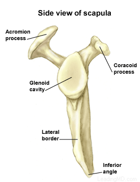
The shoulder is the most mobile joint in the human body, with a complex arrangement of structures working together to provide the movement necessary for daily life. Unfortunately, this great mobility comes at the expense of stability. Several bones and a network of soft tissue structures (ligaments, tendons, and muscles), work together to produce shoulder movement. They interact to keep the joint in place while it moves through extreme ranges of motion. Each of these structures makes an important contribution to shoulder movement and stability. Certain work or sports activities can put great demands upon the shoulder, and injury can occur when the limits of movement are exceeded and/or the individual structures are overloaded.

Click here to read more about shoulder structure

What is impingement syndrome?

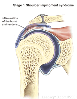
Shoulder impingement syndrome occurs when the tendons of the rotator cuff and the subacromial bursa are pinched in the narrow space beneath the acromion. This causes the tendons and bursa to become inflamed and swollen. This pinching is worse when the arm is raised away from the side of the body. Impingement may develop over time as a result of a minor injury, or as a result of repetitive motions that lead to inflammation in the bursa.

Particular shapes of the acromion may make certain individuals more susceptible to impingement problems between the acromion and the bursa. With age and the onset of arthritis, the acromion may develop bone spurs that further narrow this space. Impingement caused by bone spurs on the acromion is common in older patients who participate in sports or work activities that require overhead positions. Spurs may also result if one of the ligaments in the coracoacromial arch becomes calcified.

Impingement is classified in three grades:

  • Grade I is marked by inflammation of the bursa and tendons
  • Grade II has progressive thickening and scarring of the bursa
  • Grade III occurs when rotator cuff degeneration and tears are evident

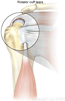
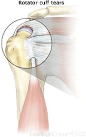
What is a rotator cuff tear?

Continual irritation to the bursa and rotator cuff tendons can lead to deterioration and tearing of the rotator cuff tendons. The tendon of the supraspinatus muscle is the most commonly involved tendon among the rotator cuff muscles. This muscle forms the top of the cuff and lies in the narrow space beneath the acromion. It is subject to the most pinching of all the rotator cuff muscles.

Rotator cuff tears can be the result of a traumatic injury or deterioration over time. Symptoms may be present, but in many cases, the patient experiences no symptoms at all. In young active people, full thickness rotator cuff tears are fairly uncommon. When they do occur, they are usually the result of a high-energy injury to the rotator cuff that is associated with throwing or overhead sporting activities. In older people, rotator cuff tears tend to be the result of wear and tear over time. Several scientific studies have shown that up to 2/3 of the population at age 70 have rotator cuff tears; many of these people had no symptoms.

Shoulder Structure

Bones and Joints

The bones of the shoulder:

  • The humerus is the upper arm bone. This is the "ball" of the shoulder's "ball and socket" joint.
  • The scapula is the flat, triangular bone commonly called the shoulder blade. Prominent areas of the scapula serve as attachment points for many muscles and ligaments.
    • The glenoid is the shallow "socket" on the side of the scapula that receives the 'ball' of the humerus. Together they form the "ball and socket" arrangement of the shoulder.
    • The scapular spine is a horizontal ridge along the back of the scapula that divides the scapula into upper and lower regions.
    • The acromion is the end of the scapular spine. It projects up to form the top of the shoulder.
    • The coracoid process is a projection towards the front of the scapula and is an attachment site for several muscles and ligaments.
  • The clavicle is the collarbone. Although it appears to be straight, it actually forms an S-shape when seen from above.
  • The thorax or rib cage, is an anchor for several muscles and ligaments. Although the ribs do not physically attach to the scapula, the thorax stabilizes and maintains proper positioning of the scapula so that the arm can function to its fullest capacity.

Additionally, there are four bone junctions, or joints:

  • The glenohumeral joint is the main joint of the shoulder. Here, the glenoid on the scapula and the head of the humerus come together. The fairly flat socket of the glenoid surrounds only 20% - 30% of the humeral head. Because of its poor fit, this joint relies heavily on the surrounding soft tissue for support. The labrum, a ring of fibrocartilage tissue, attaches to the glenoid and deepens the socket to encircle more of the humerus.
  • The acromioclavicular joint, or AC joint, is the bony point on the top of the shoulder. It stabilizes the scapula to the chest, by connecting the acromion on the scapula to the clavicle, or "collarbone". A thick disk of fibrocartilage acts as a shock absorber between the two bones. The surrounding capsule and ligaments give this joint great stability.
  • The sternoclavicular joint, or SC joint, connects the other end of the clavicle to the sternum, or "breastbone". Like the AC joint, this joint contains a fibrocartilage disk that helps the bones achieve a better fit. It also gets excellent support from its joint capsule and surrounding ligaments.
  • The scapulothoracic articulation is the area where the scapula, embedded in muscle, glides over the thoracic rib cage. The surrounding muscles and ligaments keep the scapula properly positioned so that the arm can move correctly.

Cartilage

There are two types of cartilage in the shoulder:

  • Articular cartilage is the shiny white coating that covers the end of the humeral head and lines the inside surface of the glenoid. It has two purposes:
    • To provide a smooth, slick surface for easy movement
    • To be a shock absorber and protect the underlying bone
  • Fibrocartilage is the thick tissue that forms the disks of the AC and SC joints and the labrum, the ring that deepens the glenoid. Fibrocartilage has three roles:
    • To act as a cushion in shock absorption
    • To help stabilize the joint by improving the fit of the bones
    • To act as a spacer and improve contact between the articular cartilage surfaces

Ligaments

The shoulder relies heavily on ligaments for support. Ligaments attach bone to bone and provide the "static" stability in a joint. Ligaments will alternately become tight and loose with normal motion. They keep the joint within the normal limits of movement.

  • The glenohumeral ligaments attach in layers from the glenoid labrum to form the joint capsule around the head of the humerus.
  • The coracoacromial arch is the group of ligaments that spans the bony projections of the coracoid process and the acromion.
  • The coracoclavicular ligaments and the acromioclavicular ligament provide most of the support for the AC joint.

Muscles and Tendons

Muscles and tendons work together in the shoulder to provide the "dynamic" stability of the shoulder.

There are four muscle groups in the shoulder:

  • The rotator cuff muscles are the subscapularis, the supraspinatus, the infraspinatus, and the teres minor. They are the primary stabilizers that hold the "ball" of the humerus to the glenoid "socket". The socket is too shallow to offer much security for the humerus. These four muscles form a "cuff" around the humeral head, securing it firmly in the socket. As its name implies, this group of muscles also rotates the arm. The rotator cuff protects the glenohumeral joint from dislocation, allowing the large muscles that control the shoulder to power the arm with great mobility.
  • The biceps tendon complex also helps keep the humeral head in the glenoid and helps raise the arm.
  • The scapulothoracic muscles attach the scapula to the thorax. Their main function is to stabilize the scapula to allow for proper shoulder motion.
  • The superficial muscles of the shoulder are the large, powerful outer layer of muscles that are important to the overall function of the shoulder. This group includes the deltoid muscle, which covers the rotator cuff muscles.

Bursae

A bursa is a pillow-like sac filled with a small amount of fluid. Bursae (plural) reduce friction and allow smooth gliding between two firm structures, like bone and tendon or bone and muscle. There are over 50 bursae in the human body; the largest is the subacromial bursa (under the acromion) in the shoulder. The subacromial bursa and the subdeltoid bursa (under the deltoid muscle) are often considered as one structure. This bursa separates the rotator cuff and the deltoid muscle, from the acromion.

Symptoms

What are the signs and symptoms of impingement syndrome?

Most often the onset of symptoms is related to an episode of overuse. In many patients, the episode occurred some time in the past and the shoulder has failed to return to normal. Impingement symptoms are marked by pain:

  • The pain is sharp and intermittent in its early stages.
  • As impingement progresses, the pain becomes more of a constant ache.
  • Although pain is usually present after impingement sets in, the original event that led to the problem is often relatively minor and not remembered as painful.
  • Once inflammation starts, simple movements may become painful.
  • Overhead motions tend to increase the pain. There is less space for the bursa when the arm in this position, causing more compression on the bursa.
  • Arm movements at waist level are not painful. In this position, there is more space for the bursa, and therefore it is less compressed.
  • Pain usually increases at night for two reasons. First, inflammation and swelling tend to get worse as the shoulder is used during the day, and this can lead to more pain in the evening. Second, the mind is usually less occupied in the evening, allowing pain to become a major focus of attention.

What are the signs and symptoms of a rotator cuff tear?

The symptoms of a rotator cuff tear are very similar to those of impingement syndrome with the added complaint of weakness. This weakness will vary depending on which rotator cuff tendon has been torn. For example, if the supraspinatus muscle is involved (as is most often the case) weakness will be present with forward arm elevation and overhead activity. Many patients are at first unaware of how much strength they have lost when they tear the rotator cuff.

Diagnosis

How are impingement and rotator cuff tears diagnosed?

With a careful history and physical examination, impingement and rotator cuff tears can be easily diagnosed in the doctor's office. Further testing may be necessary to determine the exact nature of a rotator cuff tear.

After the evaluation of symptoms, the doctor will perform certain muscle tests to determine whether there are tears in the rotator cuff tendons and to rule out other conditions. The doctor may place the arm in positions that reproduce the described pain to confirm the diagnosis.

There are many tests that can isolate specific areas of pain and weakness to help make the diagnosis. Further evaluation may include:

  • A diagnostic injection can help the doctor distinguish between impingement syndrome and a full thickness rotator cuff tear. A local anesthetic is injected into the inflamed bursae to eliminate the pain. If strength in the shoulder returns once the pain is blocked, it is likely that the weakness was due to pain and the rotator cuff tendons are not torn. A rotator cuff tear is suspected if strength does not return while the pain is blocked.
  • X-rays can reveal signs of arthritis, fractures, and bone spurs on the acromion. They can also reveal changes in position of the humerus and scapula that may suggest a rotator cuff tear. These images are frequently negative in the early stages of injury since X-rays show bone structure but not soft tissue.
  • An MRI (Magnetic Resonance Image) allows the physician to see muscle and other soft tissue not visible with X-ray.
  • An arthrogram is another method used to help diagnose a rotator cuff tear. A dye is injected into the shoulder and X-rays are made. If a tear in the rotator cuff tendons exists, the dye will run through the tear and make it visible on X-ray.
  • Ultrasound may also be used to diagnose a tear; however, results with this technology are difficult to evaluate and are very dependent on the skills of the technician and radiologist.
Treatment

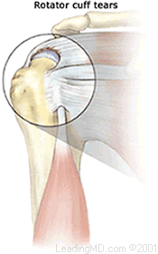
How are impingement and rotator cuff tears treated?

Impingement and rotator cuff tears can be treated non-operatively or with surgery.

Treatment for both injuries usually begins with a non-operative treatment plan. More than 2/3 of impingement patients can expect significant improvement in their symptoms with a physical therapy program alone. These results are lower in older patients and in those with large bone spurs.

When trauma causes a tear in younger patients, surgery is often the first choice of treatment. Patients with this type of injury recover best if surgery is done early. Generally, this pertains to those patients under the age of fifty with tears less than four weeks old.

Non-Operative Treatment

Non-operative treatment is similar for both impingement and rotator cuff tears. A vast majority of patients improve with this primary treatment alone. The goals of a physical therapy program include:

  • strengthening the rotator cuff tendons.
  • stretching and regaining lost motion caused by pain and inflammation.
  • allowing the humerus to be better positioned under the acromion, thus reducing compression of the bursa.

Anti-inflammatory medication may be prescribed to help reduce pain and inflammation. Many patients with rotator cuff tears can function quite well if pain and inflammation are controlled with medication and physical therapy. This is especially true for the elderly and those with low demands on the shoulder.

If symptoms have not improved with this program, the doctor may recommend a steroid injection into the bursa. Cortisone, or a similar steroid, is often combined with a local anesthetic to help control the pain and inflammation of the bursa. Steroid injections are used with caution. Damage to the rotator cuff tendons may occur with more than two or three injections over several months. Patients with diabetes are generally not good candidates for steroid injections because of problems with glucose control.

Operative Treatment

Impingement

A non-operative treatment plan is often all that is necessary for most patients with impingement syndrome. However, the small percentage of patients whose symptoms have not improved after 6 months of dedicated physical therapy may be candidates for surgery. The shoulder should be reevaluated to make sure no other problems exist.

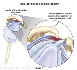
Subacromial decompression expands the space between the acromion and rotator cuff tendons. This can be done either arthroscopically or with open incisions, depending on the preference of the surgeon. During an arthroscopy, a tiny fiberoptic instrument is inserted into the joint. In many cases, the doctor can assess and repair the damage through this scope without making large incisions. Scar tissue or bone spurs can successfully be removed with either technique. If a rotator cuff tear is found at the time of surgery, it can also be repaired if necessary.

Rotator cuff tear

Not all rotator cuff tears require surgery. Many patients are content with their progress following a non-operative treatment plan. Patients who have been unable to regain lost motion and strengthen the surrounding muscles sufficiently may need a rotator cuff repair. This is often the case for the younger, more active patients who want to address continued weakness following physical therapy.

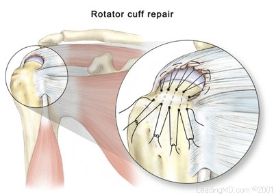
Rotator cuff repairs can be performed either arthroscopically or with open incisions. Arthroscopic techniques are new and limited to specific types of tears. An open repair that secures the rotator cuff tendons back to the humerus remains the surgical treatment of choice.

What types of complications may occur?

Complication rates after surgery are generally low. Pre-operative antibiotics are given to reduce the slight risk of infection after surgery. Infection tends to occur a little less often when arthroscopic techniques are used. Risks of major bleeding or nerve damage are extremely small. Postoperative stiffness is the major complication of both impingement and rotator cuff tears.

Recovery

Post-surgical care for impingement and rotator cuff tears are similar. General care recommendations include:

  • Incisions must be kept dry for two or three days after surgery.
  • Stitches are usually removed 7 - 10 days after surgery.

Rehabilitation programs for impingement and rotators cuff surgery differ slightly.

Impingement rehabilitation begins almost immediately:

  • Exercises to regain shoulder motion usually begin with a therapist in the first week after surgery and continue for about 6 weeks.
  • At 6 weeks, most patients have regained full motion and will continue to regain strength with a home exercise program.
  • Full recovery time after surgery varies; most patients have greatly improved at 3 months and are close to normal by 6 months.

Rotator cuff recovery is generally slower and requires more supervision. In order to achieve a full and rapid recovery, surgery should be performed as soon as the patient has full range of motion and has gained good muscle strength from a physical therapy program started when the injury is first detected. After surgery, the patient follows a closely monitored program:

  • Therapy is carefully controlled in the first 6 - 12 weeks while the tendons heal back to the bone.
  • The first goal is to regain full motion within 3 months after surgery. A therapist will assist in the early stages with gradual recovery of motion.
  • During the first 6 weeks, there is no active use of the shoulder in order to protect the surgical repair.
  • Once initial healing is achieved, a progressive stretching and strengthening program should begin.
  • Full recovery can take more than 6 months. Some patients may require more time to regain muscle strength and complete the healing process.
FAQs

Will a torn rotator cuff require surgery?

This answer to this question depends on the condition of the other shoulder muscles and the age of the patient. Many older patients have no symptoms with a rotator cuff tear and continue to function without pain or disability. In one study, up to 40% of patients over the age of 70 had no symptoms with a tear of the rotator cuff tendons. The goal of physical therapy is to maximize the function of the remaining tendons, and hopefully avoid surgery. In the younger age groups, particularly when tears are caused by a sudden injury, early surgery is generally recommended to insure a successful treatment outcome.

What is physical therapy likely to do to make mechanical impingement better?

Physical therapy is the mainstay of treatment for impingement. The vast majority of patients improve with therapy and oral anti-inflammatory medication. Strong rotator cuff muscles can relieve impingement symptoms by exerting a downward force on the humeral head, opening up the space available under the acromion. Spurs that develop beneath the acromion cannot be resolved with physical therapy, but the healthier the rotator cuff is, the less likely it is that surgery will be required.

Are there harmful effects of steroid injections for impingement?

Generally speaking, a limited number (3-5) of steroid injections into the bursa are a safe, and often effective way to locally reduce inflammation and alleviate pain. These locally applied steroids do not have the same risks associated with the chronic use of oral steroids since the body does not systemically absorb them. However, it has been shown that repeated steroid injections can damage the quality of the rotator cuff tendons if a repair is later required.

References

1. Hawkins RJ, Abrams JS. Impingement syndrome in absence of rotator cuff tears. Orthop Clin North America. 1987;3:373.

2. Lazarus MD, Chansky HA, Misra S, Williams GR, Iannoti JP. Comparison of open and arthroscopic subacromial decompression. J Shoulder Elbow Surgery. 1994;3:1-11.

3. Altchek DW, Warren RF, et al. Arthroscopic acromioplasty. Technique and results. JBJS. 1990; Sep 72(8):1198-1207.

4. Speer KP, Lohnes J, Garrett WE Jr. Arthroscopic subacromial decompression: results in advanced impingement. Arthroscopy. 1991;7(3):291-296.

5. Hawkins RH, Dunlop R. Nonoperative treatment of rotator cuff tears. COOR. 1995; Dec 321:178-188.

6. Dugas JR, Kelly BT, O'Brien, SJ. Anatomy of the Shoulder. In: Garrett WE, Speer KP, Kirkendall DT, eds. Principles and Practice of Orthopaedic Sports Medicine. Philadelphia, PA: Lippincott Williams and Wilkins; 2000:329-363.

7. Pasque CB, McGinnis, DW, Yurko-Griffin, L. Shoulder. In: Sullivan JA, Anderson SJ, eds. Care of the Young Athlete. American Academy of Orthopaedic Surgeons and American Academy of Pediatrics; 2000;323-327.

8. Thompson, WO, Warren RF, Barnes, RP, Hunt S. Shoulder Injuries. In: Schenck, Jr, RC ed. Athletic Training and Sports Medicine. 3rd edition. Rosemont, IL: American Academy of Orthopaedic Surgeons; 1999:220-232.

Accredited Business.  Rating A+  Click for Review