Shoulder Instability

Overview

Shoulder instability develops in two different ways: traumatic (injury related) onset or atraumatic onset. Understanding the differences is essential in choosing the best course of treatment. Generally speaking, traumatic onset instability begins when an injury causes a shoulder to develop recurrent (repeated) dislocations. The patient with atraumatic instability has general laxity (looseness) in the joint that eventually causes the shoulder to become unstable.

Traumatic shoulder instability is most common in young, athletic people. The younger and more active the patient is when the first dislocation occurs, the more likely it is that recurrent instability will develop. For example, if the first dislocation occurs during the teenage years, there is a 70% chance that recurrent instability will develop. However, people over 40 with a first dislocation have less than a 10% risk of developing chronic instability. Treatment strategies should be designed to suit each patient’s age and lifestyle.

What does the inside of the shoulder look like?

The shoulder is the most mobile joint in the human body, with a complex arrangement of structures working together to provide the movement necessary for daily life. Unfortunately, this great mobility comes at the expense of stability. Several bones and a network of soft tissue structures (ligaments, tendons, and muscles), work together to produce shoulder movement. They interact to keep the joint in place while it moves through extreme ranges of motion. Each of these structures makes an important contribution to shoulder movement and stability. Certain work or sports activities can put great demands upon the shoulder, and injury can occur when the limits of movement are exceeded and/or the individual structures are overloaded. Click here to read more about shoulder structure

What is traumatic shoulder instability?

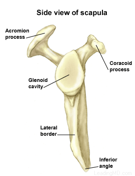
Traumatic shoulder instability begins with a first dislocation that injures the supporting ligaments of the shoulder. The glenoid (the socket of the shoulder) is a relatively flat surface that is deepened slightly by the labrum, a cartilage cup that surrounds part of the head of the humerus. The labrum acts as a bumper to keep the humeral head firmly in place in the glenoid. More importantly, the labrum is the attachment point for ligaments stabilizing the shoulder. When the labrum is torn from the glenoid, the support of these ligaments is lost. The development of recurrent instability depends upon the type and amount of damage that is done to the labrum and the supporting ligaments.

The most common dislocation that leads to traumatic instability is in the anterior (forward) and inferior (downward) direction. A fall on an outstretched arm that is forced overhead, a direct blow on the shoulder, or a forced external rotation of the arm are frequent causes of this type of dislocation. Much less common is a posterior (backward) dislocation, which is usually related to a seizure disorder or electrocution, events in which the muscular forces of the shoulder cause the dislocation.

Shoulder Structure

Bones and Joints

The bones of the shoulder:

  • The humerus is the upper arm bone. This is the "ball" of the shoulder's "ball and socket" joint.
  • The scapula is the flat, triangular bone commonly called the shoulder blade. Prominent areas of the scapula serve as attachment points for many muscles and ligaments.
    • The glenoid is the shallow "socket" on the side of the scapula that receives the 'ball' of the humerus. Together they form the "ball and socket" arrangement of the shoulder.
    • The scapular spine is a horizontal ridge along the back of the scapula that divides the scapula into upper and lower regions.
    • The acromion is the end of the scapular spine. It projects up to form the top of the shoulder.
    • The coracoid process is a projection towards the front of the scapula and is an attachment site for several muscles and ligaments.
  • The clavicle is the collarbone. Although it appears to be straight, it actually forms an S-shape when seen from above.
  • The thorax or rib cage, is an anchor for several muscles and ligaments. Although the ribs do not physically attach to the scapula, the thorax stabilizes and maintains proper positioning of the scapula so that the arm can function to its fullest capacity.

Additionally, there are four bone junctions, or joints:

  • The glenohumeral joint is the main joint of the shoulder. Here, the glenoid on the scapula and the head of the humerus come together. The fairly flat socket of the glenoid surrounds only 20% - 30% of the humeral head. Because of its poor fit, this joint relies heavily on the surrounding soft tissue for support. The labrum, a ring of fibrocartilage tissue, attaches to the glenoid and deepens the socket to encircle more of the humerus.
  • The acromioclavicular joint, or AC joint, is the bony point on the top of the shoulder. It stabilizes the scapula to the chest, by connecting the acromion on the scapula to the clavicle, or "collarbone". A thick disk of fibrocartilage acts as a shock absorber between the two bones. The surrounding capsule and ligaments give this joint great stability.
  • The sternoclavicular joint, or SC joint, connects the other end of the clavicle to the sternum, or "breastbone". Like the AC joint, this joint contains a fibrocartilage disk that helps the bones achieve a better fit. It also gets excellent support from its joint capsule and surrounding ligaments.
  • The scapulothoracic articulation is the area where the scapula, embedded in muscle, glides over the thoracic rib cage. The surrounding muscles and ligaments keep the scapula properly positioned so that the arm can move correctly.

Cartilage

There are two types of cartilage in the shoulder:

  • Articular cartilage is the shiny white coating that covers the end of the humeral head and lines the inside surface of the glenoid. It has two purposes:
    • To provide a smooth, slick surface for easy movement
    • To be a shock absorber and protect the underlying bone
  • Fibrocartilage is the thick tissue that forms the disks of the AC and SC joints and the labrum, the ring that deepens the glenoid. Fibrocartilage has three roles:
    • To act as a cushion in shock absorption
    • To help stabilize the joint by improving the fit of the bones
    • To act as a spacer and improve contact between the articular cartilage surfaces

Ligaments

The shoulder relies heavily on ligaments for support. Ligaments attach bone to bone and provide the "static" stability in a joint. Ligaments will alternately become tight and loose with normal motion. They keep the joint within the normal limits of movement.

  • The glenohumeral ligaments attach in layers from the glenoid labrum to form the joint capsule around the head of the humerus.
  • The coracoacromial arch is the group of ligaments that spans the bony projections of the coracoid process and the acromion.
  • The coracoclavicular ligaments and the acromioclavicular ligament provide most of the support for the AC joint.

Muscles and Tendons

Muscles and tendons work together in the shoulder to provide the "dynamic" stability of the shoulder.

There are four muscle groups in the shoulder:

  • The rotator cuff muscles are the subscapularis, the supraspinatus, the infraspinatus, and the teres minor. They are the primary stabilizers that hold the "ball" of the humerus to the glenoid "socket". The socket is too shallow to offer much security for the humerus. These four muscles form a "cuff" around the humeral head, securing it firmly in the socket. As its name implies, this group of muscles also rotates the arm. The rotator cuff protects the glenohumeral joint from dislocation, allowing the large muscles that control the shoulder to power the arm with great mobility.
  • The biceps tendon complex also helps keep the humeral head in the glenoid and helps raise the arm.
  • The scapulothoracic muscles attach the scapula to the thorax. Their main function is to stabilize the scapula to allow for proper shoulder motion.
  • The superficial muscles of the shoulder are the large, powerful outer layer of muscles that are important to the overall function of the shoulder. This group includes the deltoid muscle, which covers the rotator cuff muscles.

Bursae

A bursa is a pillow-like sac filled with a small amount of fluid. Bursae (plural) reduce friction and allow smooth gliding between two firm structures, like bone and tendon or bone and muscle. There are over 50 bursae in the human body; the largest is the subacromial bursa (under the acromion) in the shoulder. The subacromial bursa and the subdeltoid bursa (under the deltoid muscle) are often considered as one structure. This bursa separates the rotator cuff and the deltoid muscle, from the acromion.

Symptoms

What are the signs and symptoms of a dislocation?

If the shoulder is dislocated, it is usually very apparent:

  • The shoulder is quite painful.
  • Motion is severely restricted.
  • The shoulder appears to hang down and forward, with a large dimple evident under the acromion (in the area of the collar bone).
  • The humeral head may be visible as a bump on the front of the shoulder, or in the armpit.

To return the dislocated arm to its socket (called a reduction) usually requires a visit to the emergency department, where expert assistance can be found. Some individuals with recurrent dislocations eventually become experienced at reducing the arm themselves.

Diagnosis

How is a dislocation and traumatic shoulder instability diagnosed?

As a rule, a sudden dislocation is quite evident. The patient usually holds the arm against the side, since any attempts at motion cause pain. A large crease under the acromion and a bulge in the armpit are clues to the direction of the dislocation. However, when the shoulder spontaneously relocates into its proper position, the diagnosis can be more difficult. Patients may only report the feeling of having the shoulder "slip" before the spontaneous reduction occurred.

A qualified individual usually can relocate the humerus at the site of the injury occurrence. Once the reduction is performed, there is immediate pain relief. Without medications, some patients may be unable to relax the shoulder muscles enough to allow the reduction to take place. Often, these patients must go to the emergency department to get the reduction accomplished.

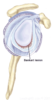
  • X-rays are usually taken to confirm the dislocation, its direction, and to check for a related fracture. After the reduction, follow up X-rays will confirm proper positioning and assess any other injuries. X-rays may reveal a "bony Bankart", which is a fracture of the anterior-inferior glenoid (front, lower portion of the glenoid). The presence of this fracture indicates that the labrum and ligaments in the front part of the shoulder are no longer attached to the glenoid.
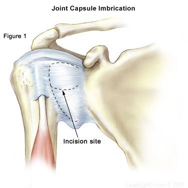
  • If X-rays do not reveal such a fracture, an MRI or arthrogram may be ordered. In this diagnostic test, the status of the labrum and ligaments can be assessed. A Bankart lesion (detachment of the anterior-inferior portion of the labrum from the glenoid) is the most common cause of recurrent instability after an injury.
Treatment

How is a dislocation and traumatic shoulder instability treated?

The initial reduction of a dislocation can be quite difficult. Contractions of the shoulder muscles can trap the humeral head against the glenoid. Gentle traction, and at times, medication may be needed to accomplish the reduction. Once the shoulder is reduced, a sling is used for a few days to protect it, and relieve discomfort. Physical therapy may help the patient regain motion in the joint.

Non-Operative Treatment

Initial treatment for recurrent instability of the shoulder centers on physical therapy. Strengthening the rotator cuff muscles and periscapular muscles (those around the scapula) gives stability to the joint. The goal of physical therapy is to help the muscles provide stability to the shoulder that the torn ligaments can no longer supply. The therapy for recurrent instability should be carefully designed for each patient since this condition often causes apprehension about certain arm positions or exercise maneuvers. Very often, physical therapy can help regain lost motion, reduce apprehension, and restore shoulder function.

Operative Treatment

Surgery is usually recommended if recurrent instability cannot be controlled with physical therapy and activity modification. The goal of surgery is to return stability to the shoulder with the least loss of motion. All shoulder procedures designed to stabilize the shoulder involve some loss of motion. The current procedures for anterior shoulder instability attempt to restore the normal anatomy without over tightening the ligaments. In certain instances, such as in young persons who have a higher risk of re-dislocation and in contact athletes who plan on continuing to participate in sports that put their shoulders at risk, surgery may be performed after the first dislocation. 

Open Labral Repair

Currently, the preferred procedure for anterior instability is an open labral repair with an anterior capsular shift. This procedure is performed through a two to three inch incision on the front of the shoulder. The torn labrum is repaired and the stretched-out anterior shoulder capsule is imbricated (overlapped) to make it smaller. This procedure is successful approximately 95% of the time in eliminating recurrent dislocations.

Arthroscopic Techniques

Recently, arthroscopic procedures such as Bankart repair have been used to repair the torn labrum and reduce capsular laxity. Arthroscopic techniques are approximately 80% successful. These procedures are performed with visualization through a small fiberoptic scope. Instruments are inserted into the joint through two or three small incisions to repair the labrum. The surgical technique is similar to the one used in an open repair. A loose capsule is more difficult to address arthroscopically. Procedures using thermal energy to shrink the loose capsule have been developed, and are still being evaluated. 

What types of complications may occur?

The major complications of anterior stabilization techniques are recurrent instability and/or loss of motion. The rate of recurrent instability depends largely on the technique used for the repair. The loss of motion can be severe, and is a function of over tightening the anterior capsule. In general, the operative shoulder should lose no more than ten degrees of external rotation. Other small risks (less than 1%) include infection, post-operative stiffness, nerve damage, or blood vessel injury.

Recovery

Non-Operative Recovery

  • Patients who have a first dislocation, and do not develop recurrent instability, will often regain full motion from a four to six week course of physical therapy.
  • Patients who do develop recurrent instability have a longer rehabilitation course and should concentrate on strengthening the shoulder muscles. Daily exercises in a home program may be recommended to help prevent instability events.

Operative Recovery

Following either arthroscopic or open operative repair and stabilization:

  • The patient will usually wear a sling for the first four to six weeks. This immobilization protects the repaired labrum while it heals to the glenoid. Until the ligaments heal, the repair must depend on the sutures used to secure the labrum.
  • During this immobilization period, elbow and wrist motion are maintained with gentle range of motion exercises.
  • Once the initial healing process is complete, physical therapy may begin. Exercises stressing range of motion are done for approximately eight weeks after surgery, or until full strength is regained.
  • Overhead sports, such as baseball or tennis, may resume about three months after surgery.
  • Contact sports are restricted for six months.
FAQs

A teenager has a first dislocation event. What is the likelihood that recurrent instability will develop?

Older research suggests that up to 90% of teenagers will develop recurrent instability after a first dislocation. More recent studies put that number closer to 70%. It is clear, however, that the younger a person is when the first injury occurs, the greater the risk is that recurrent instability will develop.

What is the point of doing physical therapy for the rotator cuff if the labrum and ligaments are torn?

The rotator cuff muscles and the periscapular muscles are important in maintaining shoulder stability. The more ineffective the ligaments are at supporting the shoulder, the more important muscular strength becomes for the control of the shoulder. Strengthening the muscles around the shoulder may provide enough stability to prevent recurrent dislocations and eliminate the need for surgery.

Should an open or arthroscopic surgery be performed?

The decision to have an open or arthroscopic repair depends on many factors. The cause of the instability, the total number of dislocations, and which technique the surgeon uses are important considerations when choosing the method of reconstruction. A thorough discussion with the surgeon of the treatment options is essential. Regardless of the technique used, the rehabilitation following surgery is the same.

Is a laser used to make the shoulder more stable?

Thermal capsulorraphy (heat treatment of the shoulder capsule) is a new technique developed to achieve shoulder stability. Newer techniques involve a radio frequency probe instead of a laser to shrink the capsule. This type of treatment continues to evolve, and its results are still being evaluated.

References

1. Hovelius L, et al. Primary anterior dislocation of the shoulder in young patients. A ten-year prospective study. JBJS. 1996:78A:1677-1684.

2. Rockwood C, et al. The capsular imbrication procedure for recurrent anterior instability of the shoulder. JBJS. 1996;78A:246-259.

3. Gartsman GM, et al. Arthroscopic treatment of anterior inferior glenohumeral instability. A two to five year follow up. JBJS. July 2000;82A:991-998.

Accredited Business.  Rating A+  Click for Review